Boucharaba et al. 10.1073/pnas.0600979103. |
Supporting Figure 8
Supporting Figure 9
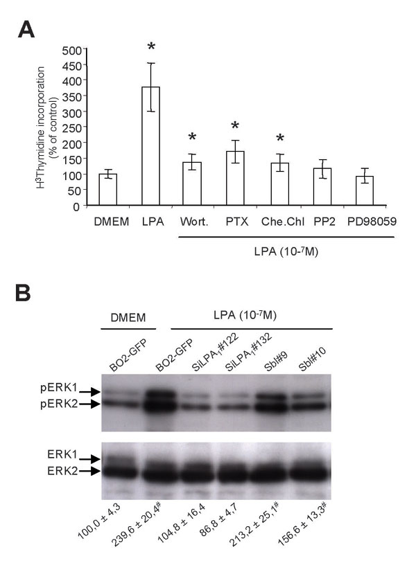
Fig. 8. Lysophosphatidic acid (LPA) stimulates MDA-BO2/GFP cell proliferation through a LPA1-dependent activation of mitogen-activated protein kinase (MAPKinase) ERK1/2. (A) Quiescent MDA-BO2/GFP cells plated in 96-well tissue culture plates were incubated with or without Wortmannin (10-7 M), chelerythrine chloride (10-6 M), PP2 (10-7 M), or PD98059 (10-5 M) 8 h before LPA (10-7 M) stimulation for 16 h in serum-free medium (DMEM) containing 0.1% (wt/vol) BSA fatty acid free and then pulsed with [H3]thymidine for 8 h. [H3]thymidine incorporation was quantified and data expressed as percent of MDA-BO2/GFP cells treated with DMEM are the mean ± SD of five replicates and are representative of at least three separate experiments. *P < 0.001, treated cells versus control cells. (B) Quiescent MDA-BO2/GFP cells and transfectants were treated without (DMEM) or with LPA (10-7 M) for 2 min. Cells were then lysed in the presence of NA3VO4 and protease inhibitors. Then, 20 mg of total protein extract was resolved on 10% SDS/PAGE and transferred onto poly(vinylidene difluoride) (PVDF) membrane. Immunodetection of phosphorylated ERK1/2 (Upper) and total ERK1/2 (Lower) were achieved by using specific polyclonal antibodies (Santa Cruz Biotechnology). The intensity of immunoreactive bands was then quantified by using the computerized image analysis system calculated in each condition. Data indicated at the bottom are expressed as percent of MDA-BO2/GFP treated with DMEM (mean ± SD of three independent experiments). #, P < 0.05, treated cells versus control cells.

Fig. 9. Effect of LPA on MDA-BO2/GFP tumor cell secretion of cytokines, chemokines, and growth factors. Conditioned media of MDA-BO2/GFP cells treated without (unstimulated) or with 1-oleoyl LPA (1 mM) were harvested after 48 h. Membranes of RayBio Human Cytokine Antibody Array V (RayBiotech, Norcross, GA) were blocked with the saturated buffer and then incubated with the conditioned medium (1 ml) from 106 cells. After washing, membranes were incubated with a mixture of 79 biotinylated antibodies. Then, membranes were washed and incubated with a peroxidase-labeled streptavidin solution. Detection of immunoreactive spots was carried out by using the enhanced chemiluminescence (ECL) detection system. Cytokines, growth factors, and chemokines secreted under unstimulated conditions were indicated by arrows (Left). Cytokines, growth factors, and chemokines up-regulated in the presence of LPA are indicated by arrowheads (Right). (Pos, positive control; Neg, negative control).