Hinrichsen et al. 10.1073/pnas.0600312103. |
Supporting Figure 6
Supporting Figure 7
Supporting Figure 8

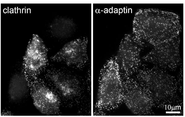
Fig. 6. AP2 membrane domains persist after clathrin depletion by RNA interference. HeLa cells were transfected with oligo I from ref. 1 and processed for immunofluorescence 3 days later.
1. Hinrichsen, L., Harborth, J., Andrees, L., Weber, K. & Ungewickell, E. J. (2003) J. Biol. Chem. 278, 45160-45170.

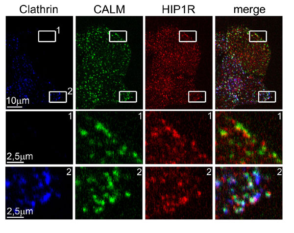
Fig. 7. Hip1R staining overlaps with that of CALM in control and clathrin-depleted cells. HeLa cells were transfected with oligo I from ref. 1, transfected with the same oligo after 48 h again, and processed for immunofluorescence after another 48 h. For triple-labeling fixed and permeabilized cells were incubated first with goat anti-CALM for 1 h at 37°C and then with fluorescine-labeled donkey anti-goat antibody. For blocking the anti-goat secondary antibody the cells were fixed again with 4% paraformaldehyde in PBS for 10 min and incubated for 5 min with TBS and subsequently for 10 min in 10% goat serum. The cells were then fixed again in paraformaldehyde for another 10 min and then incubated for 5 min with TBS. Subsequently the cells were reacted with primary antibodies against clathrin (rabbit serum R461) and Hip1R (mouse monoclonal) for 1 h at 37°C and finally with the secondary antibodies goat anti-rabbit, Alexa Fluor 633-, and rhodamine-conjugated anti-mouse antibodies (Molecular Probes).
1. Hinrichsen, L., Harborth, J., Andrees, L., Weber, K. & Ungewickell, E. J. (2003) J. Biol. Chem. 278, 45160-45170.

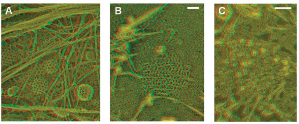
Fig. 8. Anaglyph stereo views of the inner surface of the plasma membrane of "unroofed" HeLa cells. (A) Plasma membrane of control cell. (B) Plasma membrane of clathrin-depleted cell. (C) ImmunoGold (10 nm) labeling of AP2 after clathrin depletion. Note the flat appearance of the AP2 membrane domain. (Scale bar: 100 nm.)