Supporting information for Groves (2003) Proc. Natl. Acad. Sci. USA, 10.1073/pnas.0830019100

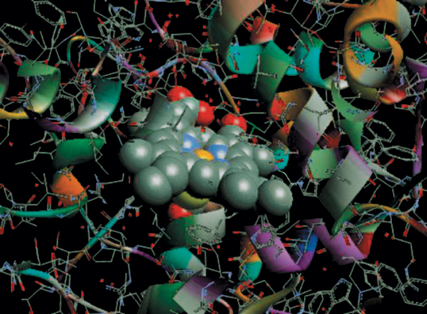
Fig. 5.
Crystal structure of the active site of chloroperoxidase (EC 1.11.1.10) from C. fumago. Protein framework is shown as ribbons. The heme is buried in a hydrophobic binding pocket containing the iron-coordinating cysteinate ligand (yellow). Adapted from the x-ray atomic coordinates of CPO (1).1. Sundaramoorty, M., Terner, J. & Poulos, T. L. (1995) Structure (London) 3, 1367–1377.