Supporting information for Korth et al. (2003) Proc. Natl. Acad. Sci. USA, 10.1073/pnas.2627989100

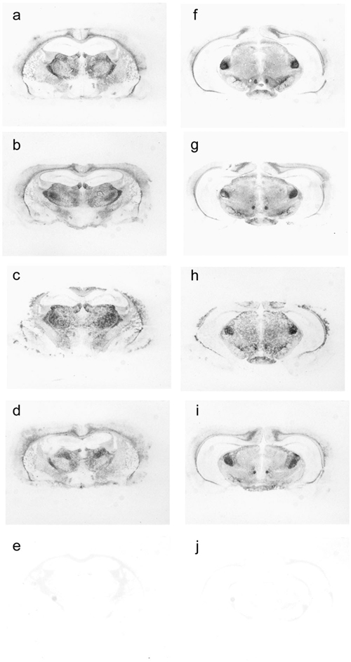
Fig. 6.
Histoblots of Tg22372 mice inoculated with different cases of with sporadic Creutzfeldt–Jakob disease (sCJD) (MM1). Coronal sections through the thalamic-hippocampal area (a–e) or midbrain area (f–j) are depicted. (a and f) sCJD(MM1) (EC). (b and g) sCJD(MV1) (WP). (c and h) sCJD(MM1) (RG). (d and i) Second passage of sCJD(MM1) (RG). (e and j) Uninoculated control. The histoblots show a similar PrPSc deposition pattern for all inoculated mice.