Supporting information for Korth et al. (2003) Proc. Natl. Acad. Sci. USA, 10.1073/pnas.2627989100

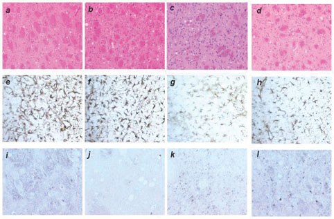
Fig. 8.
Neuropathology of Tg22372 mice inoculated with different cases of sporadic Creutzfeldt–Jakob disease (sCJD) (MM1). Columns correspond to different inocula: (a, e, i) sCJD(MM1) (EC); (b, f, and j) sCJD(MV1) (WP); (c, g, and k) sCJD(MM1) (RG); (d, h, and l) second passage of sCJD(MM1) (RG). Rows correspond to different staining techniques: hematoxylin and eosin (Top); α-GFAP immunostaining (Middle); or α-PrP immunostaining (Bottom). The caudate nucleus is depicted, at 25´ objective. The neuropathological lesion profile after first passage of sCJD shows minimal differences in the degree of vacuolation (compare a, b, and c), intensity of astrocytic gliosis (compare e, f, and g), and PrP immunostaining after hydrolytic autoclaving (compare i, j, and k). Furthermore, there is no change in the neuropathological lesion profile after second passage (compare d, h, and l with the others).