Canty et al. 10.1073/pnas.0607350103. |

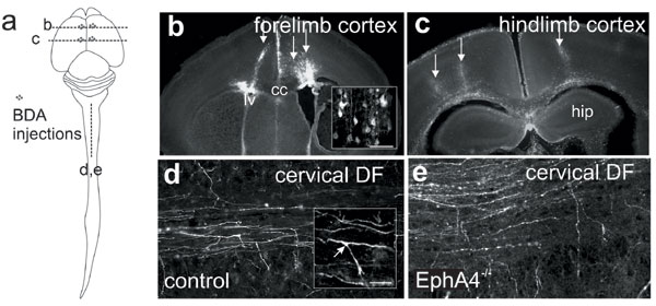
Fig. 5. Anterograde tracing of CST. Location of injection sites for the fore- and hindlimb motor cortex (arrows) are shown schematically in a, and representative coronal sections are shown in b and c. Landmarks, including the lateral ventricles (lv), corpus callosum (cc), and the rostral hippocampal commissure (hip), were used to identify different regions of the motor cortex. Uptake of BDA into the layer 5 pyramidal cells (b Inset) allowed visualization of axons in horizontal or sagittal sections in the dorsal funiculus (d and e) and identification of branch points (arrow, d Inset). (Scale bars: d and e, 100 mm; b and d Insets, 50 mm).

Fig. 6. EphA4 expression in postnatal dorsal funiculus and motor cortex. (a-f) EphA4 (green) is expressed in the DF of the cervical and lumbar enlargements at P0, P5, and P11. Dotted lines indicate the boundary of the dorsal funiculus. Arrowheads indicate the midline of the spinal cord. (g-g¢¢) Horizontal view of ventral DF labeled for EphA4 (g) with BDA-labeled CST axons (g¢, red) and merge of EphA4 and BDA labeling showing colocalization (g¢¢). (h-j) Adjacent sections of P5 forelimb motor cortex are shown, stained with thionin (h) and EphA4 (i), as well as a section through P5 hindlimb motor cortex labeled with EphA4 (j). Dotted lines in h and i indicate the area containing layer 5 neurons in the motor cortex. (Scale bars: c and d, 20 mm; a, b, g-g¢¢, e, and f, 50 mm; h-j, 100 mm.)

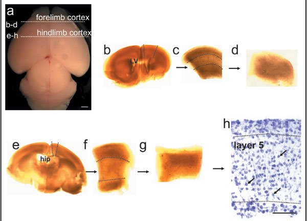
Fig. 7. Isolation of layer 5 of the fore- and hindlimb motor cortex for in vitro assay. (a-g) The P3 brain was cut into thick coronal sections in sterile conditions, and regions of the forelimb motor cortex (rostral to the genu of the corpus callosum and above the lateral ventricles; b and c) and caudomedial hindlimb motor cortex (rostral tip of the hippocampal commissure; e and f) were removed. The upper and lower thirds of the cortex were then dissected away to leave the middle third of the cortex, which predominantly contains the large pyramidal neurons of layer 5 (d and g). (h) The identity of these layers was confirmed by staining with thionin to visualize the large pyramidal cell bodies of layer 5 (arrows). hip, rostral tip of the hippocampal commissure; LV, lateral ventricles. (Scale bars: 100 mm.)

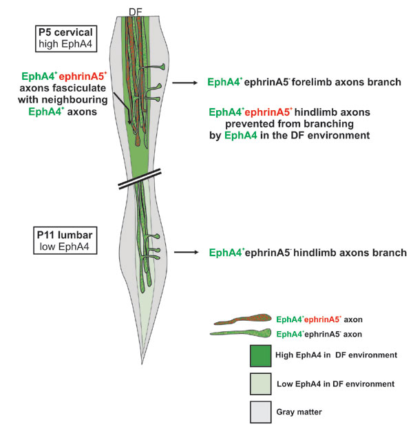
Fig. 8. Proposed mechanism of EphA4-ephrinA5 interaction in the generation of topography in the CST. At P5, forelimb axons express low levels of ephrinA5 and can branch at the cervical level into an environment containing EphA4. EphrinA5+ hindlimb axons are prevented from branching at this level. Branching could be prevented by EphA4 in the environment and/or the enhanced fasciculation of the ephrinA5+ axons with EphA4 on neighboring axons. By P11, there is a decrease of both ephrinA5 expression on hindlimb axons and EphA4 expression in the DF environment, which permits the hindlimb axons to branch out of the DF at the lumbar level.