Supplementary material for Sheppard et al. (2000) Proc. Natl. Acad. Sci. USA 97 (14), 7802-7807.

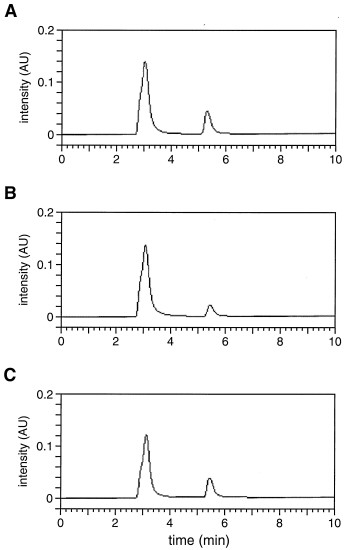
Fig. 10. HPLC chromatograms comparing authentic guanine and the nucleobase product resulting from reaction of the 10-28 DNA enzyme and 35mer DNA substrate. (A) Solution containing 1 nmol of authentic guanine/2 mM CaCl2/10 mM NaCl/4 mM Na2EDTA/10 mM Mes (pH 5.2). (B) Products of a reaction containing 0.5 nmol of enzyme and 2 nmol of substrate, were incubated in the presence of 2 mM CaCl2/10 mM NaCl/10 mM Mes (pH 5.2) at 25ºC for 20 h, and then quenched by addition of 20 mM Na2EDTA. The products were not treated with piperidine before HPLC analysis. (C) Mixture of equal amounts of the material used to generate the chromatograms shown in A and B. Absorbance was monitored at 280 nm (e = 7.6 AU<949>m mol). The solvent front produced a peak at about 3 min and guanine eluted between 5 and 6 min. No other peaks were observed on the HPLC chromatogram, monitored at either 254 or 280 nm. The guanine peak was collected and its complete UV spectrum was obtained, which was identical to that of authentic guanine.