
Magarinos et al. 10.1073/pnas.0608785103. |
Fig. 5. Effect of deep hibernation (Hib) and induced awakening (Awake, 2 and 3 h) on the number of dendritic branch points (Upper) and total dendritic length (Lower) of pyramidal neurons from layer V of parietal cortex. Bars represent mean + SEM.
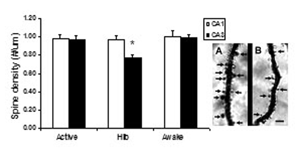
Fig. 6. Effect of deep hibernation (Hib) and induced awakening (Awake, 3 h) on spine density in apical dendrites of CA1 and CA3 pyramidal neurons. *, P < 0.05 compared with active and awake groups, one-way ANOVA, Tukey post hoc test. Bars represent means plus SEM. (Right) A photomicrograph of Golgi-impregnated apical dendrites from CA3 pyramidal neurons of active (A) or hibernating (B) European hamsters. The arrows indicate dendritic spines in the plane of focus. Note the greater density of dendritic spines in A compared with B. (Scale bar: 10 mm.)