Supplementary material for Incardona et al., Proc. Natl. Acad. Sci. USA, 10.1073/pnas.220251997

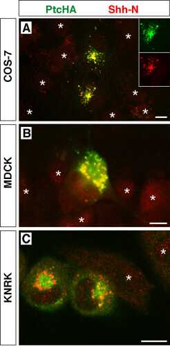
Fig. 5. Internalization of Shh-N by cells expressing PtcHA. PtcHA immunofluorescence is shown in green and Shh immunofluorescence in red; yellow or orange indicates colocalization. Untransfected cells are marked with asterisks. (A) COS-7 cells; a pair of PtcHA-transfected cells are shown among a near-confluent field of untransfected cells. PtcHA+ vesicles are typically observed peripherally in filopodia, as well as in a perinuclear cluster. (Insets) The individual single-color images for the lower cell. (B) MDCK cells; a single PtcHA-expressing cell is shown among five untransfected cells. (C) KNRK cells; two PtcHA-expressing cells are shown with two untransfected cells. PtcHA and Shh immunofluorescence is evident in perinuclear vesicles. In the red channel, all cells show background staining in nuclei and occasional autofluorescent granules, as well as some nonspecific cytoplasmic staining in untransfected cells. (Scale bars = 10 m M.)