Supplementary material for Kaech et al. (1999) Proc. Natl. Acad. Sci. USA 96 (18), 10433-10437.

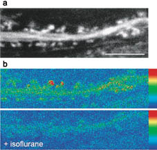
Fig. 5. Volatile anesthetics block spine motility in organized tissue. Organotypic cultures were obtained from hippocampi of transgenic animals expressing actin-GFP under the control of the chicken beta-cytoplasmic actin promoter, which in brain drives expression mainly in neurons. (a) Confocal image of the basal dendritic segment of a CA1/CA2 pyramidal cell in a slice maintained in culture for 6 weeks. (Bar = 10 mm.) (b) Isoflurane inhibits spine motility in organized tissue. To compare the extent to which spine motility was inhibited by anesthetics, changes in shape recorded before an after perfusion with isoflurane are shown as summed differences between 40 successive frames recorded 10 sec apart and displayed by using a color code scale (strip at right of images). Under control conditions (Upper), pronounced changes were associated with spine heads. In the presence of the anesthetic (Lower), these were reduced to background levels.