Supplementary material for Kaech et al. (1999) Proc. Natl. Acad. Sci. USA 96 (18), 10433-10437.

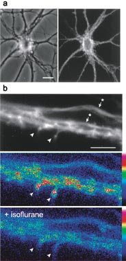
Fig. 6. Inhibition of spine motility by anesthetics monitored by using membrane-targeted GFP. (a) Primary rat hippocampal neurons transfected with a membrane-targeted GFP (see Methods in main text) develop and mature normally. (Left) Phase contrast image of a neuron maintained in culture for 3 weeks. (Right) Corresponding GFP fluorescence image. (Bar = 15 mm.) (b) Higher magnification image of a dendrite segment with spines (arrowheads) apposed on one side by segments of the transfected neuron’s own axon (arrows with stars) recursing along its dendrites. Axons of untransfected neurons were observed in phase contrast contacting the other spines. Because the membrane-targeted GFP distributes evenly throughout the cell’s plasma membrane (i.e., there is no specific enrichment of the fluorescent signal in spines as occurs with spine-targeted GFP-actin), the axon, dendrite shafts, and spines of neurons transfected with membrane-targeted GFP all show a similar level of fluorescence. (Bottom) Summed difference images from time-lapse recordings of 40 frames taken 15 sec apart. These recordings show that under control conditions (Middle), the most pronounced changes are observed at spine heads (arrowheads; compare fluorescence image, Upper, with summed difference images, Middle) and that these are practically eliminated when isoflurane is present (Bottom). Note that the absence of a signal above background over the spine head does not reflect retraction of the spine, but does reflect the suppression of motility that would have generated a difference image signal between successive frames. (Bar = 10 mm.)