Supplementary material for Wang et al. (1999) Proc. Natl. Acad. Sci. USA 96 (21), 12150-12155.

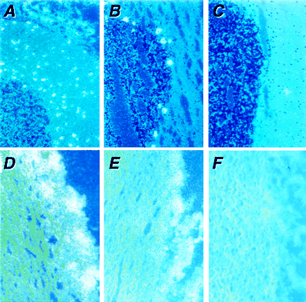
Fig. 7.
Localization of nNOS exon 1 variants in cerebellum and aorta by using in situ hybridization. Representative serial sections from normal baboon cerebellum (A–C) and human aorta (D–F) were hybridized with 35S-labeled antisense riboprobes against exon 1f (A and D), exon 1g (B and E), or exon 1a (C and F). Serial sections hybridized with a corresponding sense probes did not show hybridization (data not shown). Autoradiographs were photographed by using a combination of bright-field illumination and polarized epiillumination. In cerebellum, exons 1f and 1g were differentially expressed in the cerebellar granular layer or Purkinje cells, respectively. Exon 1a was not detected in cerebellum. Exons 1f and 1g were colocalized in the perivascular nerves of the aorta, whereas exon 1a was not detected.