Woo Lim et al10.1073/pnas.0611123104XXYYYYY103. |

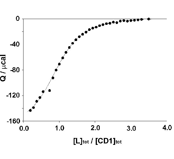
Fig. 10. Isothermal titration calorimetry of guest L to host vesicles of CD1. [L] = 5.0 mM and [CD1] = 0.5 mM in H2O at pH 9. T = 303 K.
Fig. 11. (A) Size distribution of vesicles of CD1 (60 mM) according to dynamic light scattering. Black, vesicles; red, vesicles and [CuL2] = 60 mM; green, vesicles and [CuL2] = 125 mM. (B) Size distribution of vesicles of CD1 (33 mM) according to dynamic light scattering. Black, vesicles; red, vesicles and [NiL3] = 1.0 mM; green, vesicles and [NiL3] = 1.0 mM and [EDTA] = 5.0 mM. pH = 9.0.
Fig. 12. Optical density at 400 nm of a solution of vesicles of CD1 (10 mM) in the presence of CuL2 (0.25 mM) (closed circles), NiL3 (50 mM) (open circles), NiL3 (50 mM) and bCD (10 mM) (closed squares), NiL3 (50 mM) and bCD (0.1 mM) (closed triangles), and NiL3 (50 mM) and bCD (1 mM) (open triangles). pH = 9.0.