Supplementary material for Minasov et al. (2000) Proc. Natl. Acad. Sci. USA 97 (12), 6328-6333.

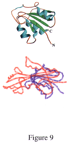
Fig. 9.
Structural comparison between Maf and a-D-glucose-1,6-bisphosphate phosphoglucomutase (PMG). Secondary structure of PMG (Left) and structural comparison between Maf and PMG (red and blue, respectively; Right).