
Ravindran et al. 10.1073/pnas.0701363104. |
Fig. 7. Blood phagocytes are recruited to the site of infection but do not participate directly in antigen presentation. (A) C57BL/6 mice were left uninfected (naïve) or were infected s.c. with 2 ´105Salmonella-EaRFP s.c. The injection site was removed and a cell suspension prepared 12 and 24 h later and analyzed for the expression of Gr-1 and CD11b. (B) WT and CCR6-deficient mice were adoptively transferred with 2 ´ 106 CFSE-labeled TEa T cells and infected s.c. with 2 ´ 105Salmonella-EaRFP or left uninfected (Transfer Only). Some mice were injected with purified Gr1+CD11b-, Gr1+CD11b+, Gr1-CD11b- blood monocytes, or blood CD4 cells from WT or MHC class-II-deficient mice, 10 min before infection. Three days after infection, cervical lymph were recovered and stained with antibodies to CD4 and CD90.1 to identify TEa T cells. Plots show CFSE staining after gating on TEa T cells and are representative of two to three mice per group.

Fig. 8. Construction of Salmonella-RFP and Salmonella-EaRFP. Virulent Salmonella were transformed with a plasmid vector encoding EaRFP fusion protein (A) or RFP (B). After induction of protein expression, bacteria were fixed and examined by flow cytometry for red fluorescence. Filled histograms show untransformed virulent Salmonella and line histograms show bacteria expressing EaRFP or RFP.

Fig. 9. DC populations in lymph nodes of Wt and CCR6-deficient mice. Submandibular and cervical lymph nodes were harvested from Wt and CCR6-deficient mice using collagenase/EDTA, and CD11c+ cells enriched. To identify DC subsets, CD11c+ cells were stained with antibodies specific for CD4, CD8a, CD11b, B220, and Epithelial cell adhesion molecules (EpCam).
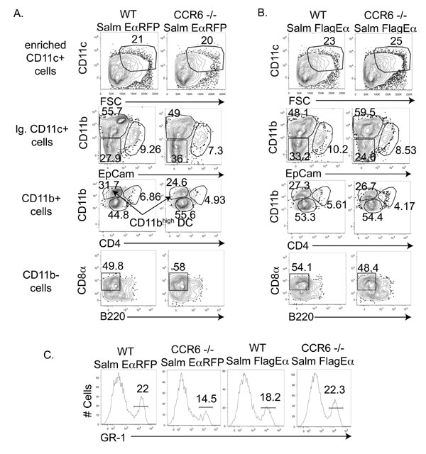
Fig. 10. DC populations in lymph nodes of Wt and CCR6-deficient mice after Salmonella infection. (A-B) Draining lymph nodes were harvested from WT or CCR6-deficient mice, 24 h after infection with Salmonella-EaRFP (A) or Salmonella-FlagEa (B), and CD11c+ cells were enriched. To identify DC subsets, CD11c+ cells were stained with antibodies specific for CD4, CD8a, CD11b, B220, and EpCam. (C) Gr-1 expression levels present on CD11bhigh DCs detected in the lymph nodes of infected mice.

Fig. 11. Expression of chemokine receptors on blood leukocyte populations. Blood leukocytes from uninfected or Salmonella-infected CCR6-GFP knock-in heterozygous mice were stained with antibodies to Gr1+ and CD11b-, and Gr1+CD11b-, Gr1+CD11b+, or Gr1-CD11b- blood cells identified as shown in boxed gates in upper plots. Expression of CCR6 was examined in each population by GFP expression and expression of CCR2 was examined by staining with specific antibodies.