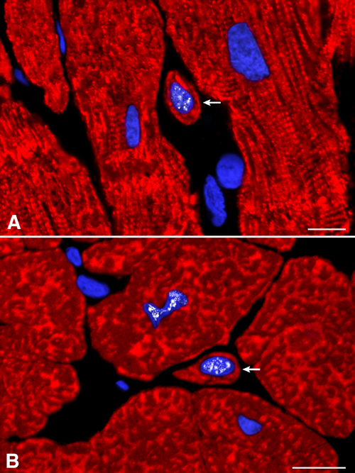
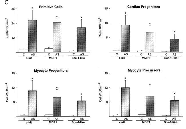
Fig. 12.
Identification of developing myocytes. (A and B) Small differentiating myocytes (arrows) negative for stem cell antigens and positive for cardiac myosin heavy chain (red fluorescence). Nuclei are labeled by MEF2 (white fluorescence). These examples correspond to hypertrophied hearts. (Scale bars = 10 mm.) (C) Numerical density of primitive and early committed cells in the myocardium. Results are mean ± SD. Asterisks indicate P < 0.001 between results in hypertrophied (AS) and control (C) hearts.