[View Larger Version of this Image]
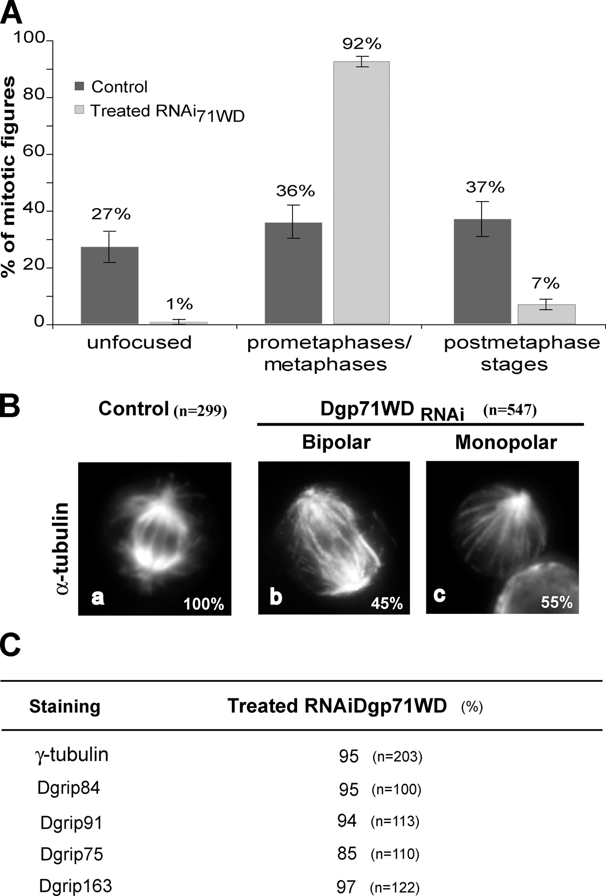
Figure S2. Phenotypes observed after Dgp71WD depletion in S2 cells. (A) Distribution of cells at the different stages of mitosis. The different mitotic stages were scored in >250 cells undergoing mitosis. Error bars represent confidence level calculated for P95. (B) Analysis of spindle morphology by immunofluorescence. Control cells or Dgp71WD-treated cells were stained for microtubules. (C) Accumulation of centrosomal components in mitotic S2 cells after depletion of Dgp71WD. The spindle poles were immunostained with antibodies against γ-tubulin, Dgrip84, Dgrip91, Dgrip75, or Dgrip163. n is the number of prometaphases/metaphases analyzed.