[View Larger Version of this Image]
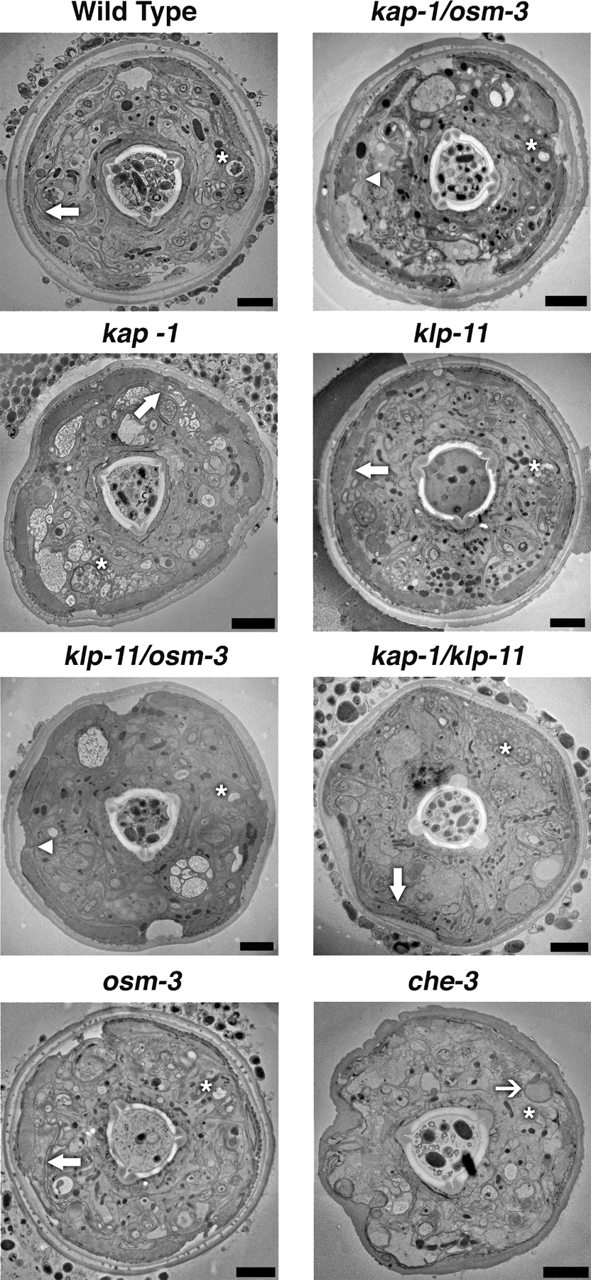
Figure S1. Transmission electron micrograph of sections of each strain ∼5 µm from the tip of the head. Within each micrograph, a single amphid pore is indicated by an asterisk, and a single AWC cilium is marked by a thick arrow if it has a normal fan-like structure, a thin arrow if the AWC cilium is present but has a changed morphology, or an arrowhead to indicate the absence of the AWC cilium. Bars, 2 µm.