[View Larger Version of this Image]
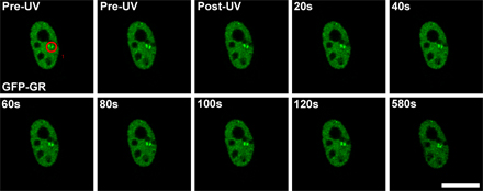
Figure S3. Changes in specific chromatin structure upon UV laser microirradiation. MMTV gene array-containing cells expressing GFP-GR localized to the nucleus were incubated in the absence of the sensitizing Hoechst dye but exposed to UV laser irradiation in specific spots within the nucleus, as shown by the red circle encompassing the MMTV gene array in the pre-UV panel. Images from subsequent post-UV irradiation time points are shown. Bar, 10 µm.