[View Larger Version of this Image]
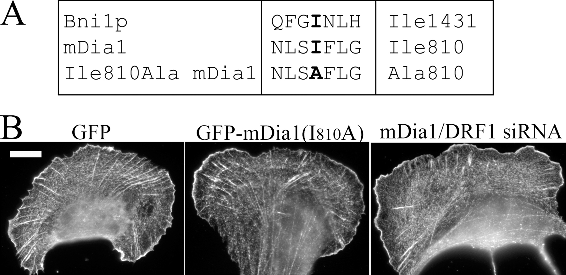
Figure S1. Overexpression of a loss-of-function mDia1/DRF1 mutant induces abnormal dorsal stress fiber morphology and accumulation of α-actinin in these structures. (A) Alignment of mDia1 and Bni1 sequences surrounding the conserved isoleucine (in bold) that is essential for actin filament nucleation/polymerization activity of the FH2 domain (Xu et al., 2004). (B) U2OS cells were transfected with GFP, GFP-mDia1(I810A), or mDia1/DRF1 siRNA; replated 20 h (plasmids) or 44 h (siRNA) after transfection; and fixed 4 h after replating. α-Actinin was visualized by anti-α-actinin antibodies. The dorsal stress fibers in GFP-transfected cells are indistinguishable from the dorsal stress fibers in wild-type cells (Fig. 4). GFP-mDia1(I810A) and mDia1/DRF1 siRNA-transfected cells exhibit short dorsal stress fibers and abnormal α-actinin accumulation into these structures. Similarity in phenotypes suggests that the effects of RNAi were specific to mDia1/DRF1 and that the actin filament nucleation/polymerization activity is crucial for the function of this protein during dorsal stress fiber formation. Bar, 10 µm.