[View Larger Version of this Image]
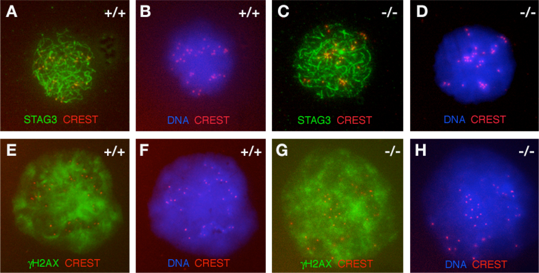
Figure S2. Distribution of STAG3 and γH2AX in wild-type and Sycp2-/- spermatocytes. Surface-spread nuclei of spermatocytes were immunostained with anti-STAG3 serum (green) or γH2AX-specific antibodies (green), CREST (red), and DNA (blue). We found no difference in the amount of γH2AX between wild-type and Sycp2-/- spermatocytes, suggesting that absence of axial elements in Sycp2-/- spermatocytes does not abolish the formation of DSBs. (A) STAG3 localization in wild type. (C) STAG3 localization in Sycp2-/- spermatocytes. (E) γH2AX staining in wild type. (G) γH2AX staining in Sycp2-/- spermatocytes. (B, D, F, and H) DAPI staining of the same cells shown in A, C, E, and G, respectively.