[View Larger Version of this Image]
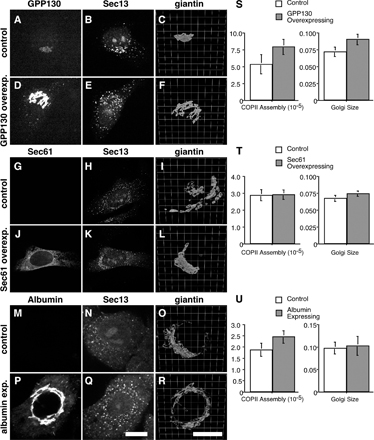
Figure S4. Golgi size is increased by GPP130 but not Sec61 or albumin expression. (A-R) HeLa cells stably transfected with GPP130-GFP (A-F), HeLa cells transiently transfected with Sec61-GFP (G-L), or HepG2 cells expressing albumin (M-R) were paraformaldehyde-fixed and analyzed to reveal GFP fluorescence (A, D, G, and J), albumin staining (M and P), and Sec13 staining (B, E, H, K, N, and Q), as well as giantin staining shown after 3D rendering (C, F, I, L, O, and R). Control cells were those that exhibited little or no detectable expression, whereas expressing cells were neighboring cells yielding strong staining. Bars, 10 µm. (S-U) COPII assembly and Golgi/cell size were quantified as described for the matched control cells and the cells expressing GPP130-GFP (S), Sec61-GFP (T), or albumin (U). Values are means (± SEM; >15 cells each).