[View Larger Version of this Image]
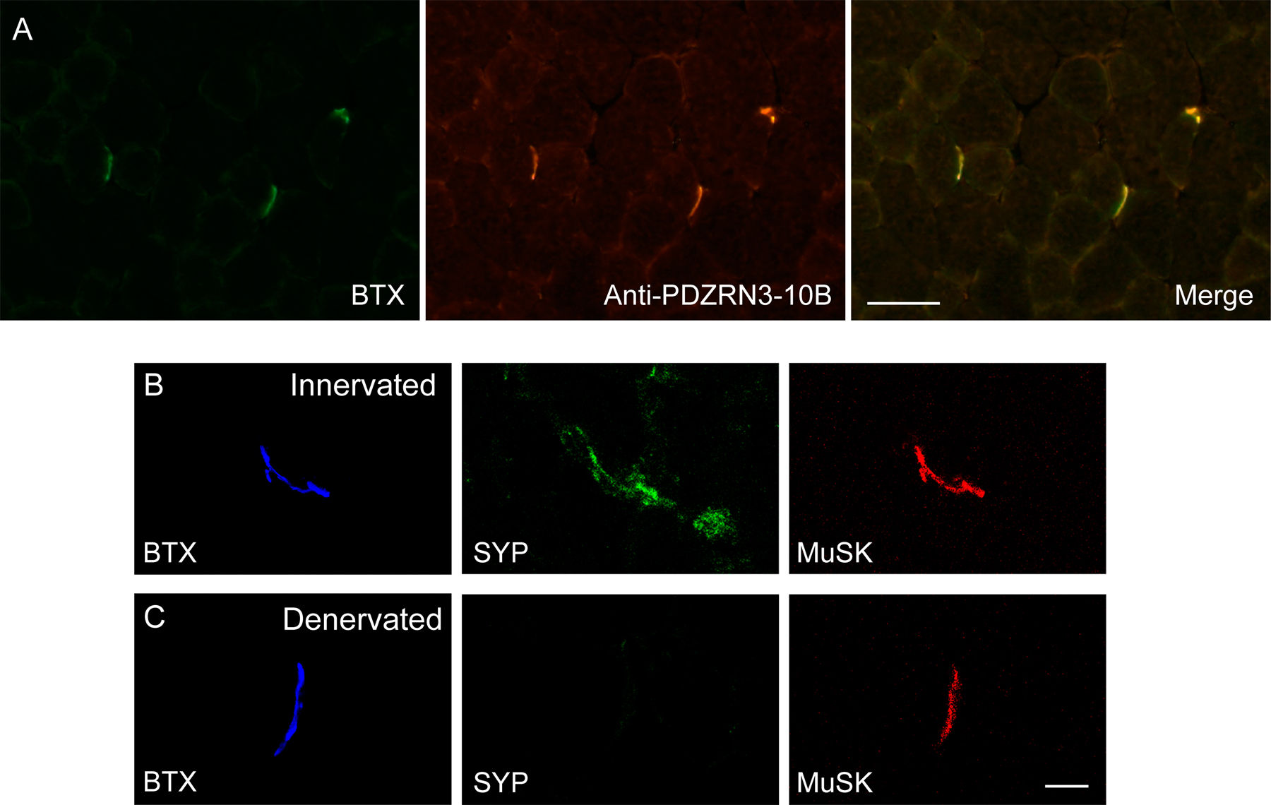
Figure S1. (A) weak staining of extrasynaptic muscle membrane by anti-PDZRN3 antibody. (B and C) MuSK staining persists after denervation.