[View Larger Version of this Image]
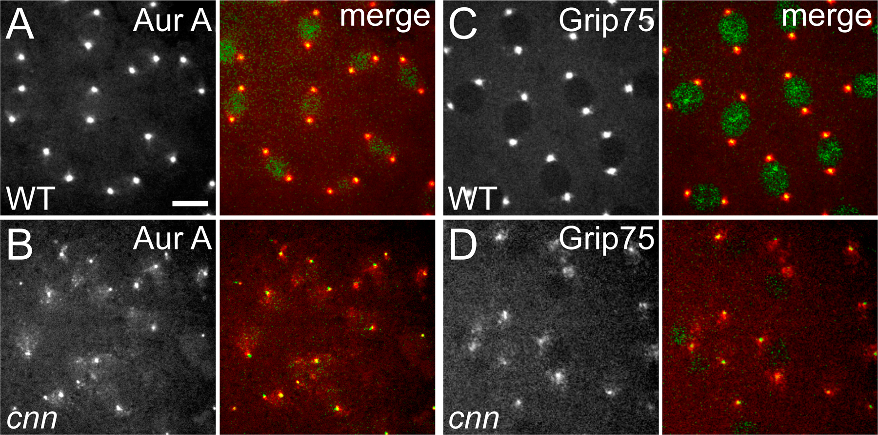
Figure S1. The centrioles in cnn embryos are associated with some PCM but are not properly centered within it. The WT and cnn embryos shown here express the PCM markers Aurora A-GFP (A and B) or Grip75-GFP (C and D; all pseudocolored red in the merged image), as well as the centriole marker mRFP-Fzr (pseudocolored green in the merged image; note that mRFP-Fzr is also concentrated in the nucleus in interphase). (A and C) In WT embryos, the centrioles are always well centered within the PCM. (B and D) In cnn embryos, the centrioles are associated with PCM, but they cannot maintain their proper connection to the PCM. Only relatively small amounts of Aurora A-GFP and Grip75-GFP are recruited to the centrioles compared with GFP-D-TACC, so only small amounts of these proteins remain associated with the centrioles as they move around the cnn embryo. See Video 2. Bar, 10 µm.