[View Larger Version of this Image]
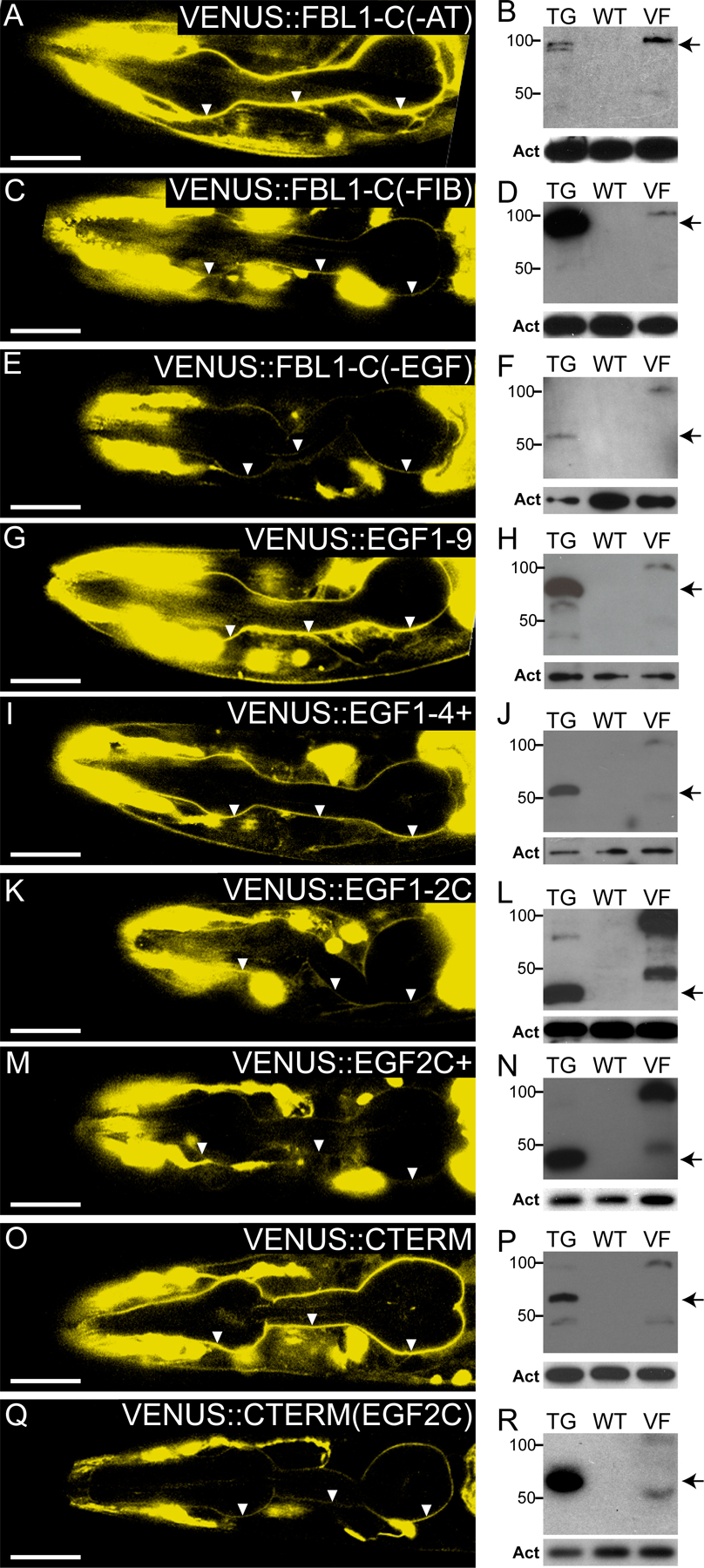
Figure S1. Localization and protein expression of FBL-1 transgenes. (left) 0.8-µm confocal slices of fbl-1(q771) transgenic L4 larvae, anterior to left; transgenes are indicated in the figure. Arrowheads indicate pharyngeal basal lamina. Bars, 20 µm. (right) Anti-VENUS Western blots. TG, extract corresponds to transgenic strain pictured on the left. WT, extract from nontransgenic wild-type strain; negative control. VF, extract from VENUS::FBL-1 strain; positive control. Act, actin loading control. Arrows mark the predicted size of transgenic proteins. All transgenes accumulated in major bands at the predicted sizes for each fragment.