[View Larger Version of this Image]
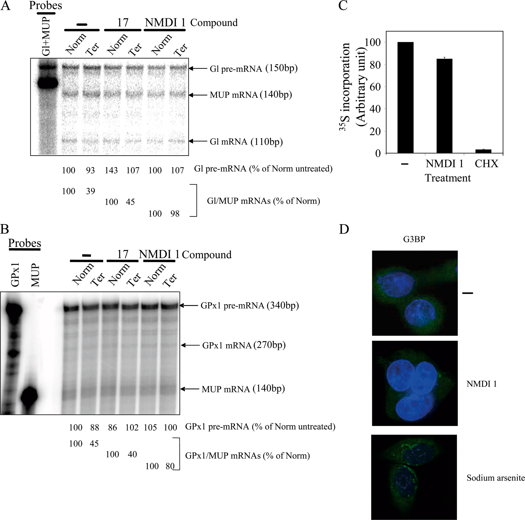
Figure S1. NMDI 1 is a specific NMD inhibitor. (A) Measure of NMD inhibition by NMDI 1 using RPA. 3 μg of total RNAs from cells treated with DMSO (−), 5 μM of compound 17 (17), or 5 μM of NMDI 1 were analyzed by RPA and loaded on 6% denaturing gel. The levels of Gl pre-mRNA and mRNA either wild type (Norm) or harboring a premature termination codon (Ter) and MUP mRNA are measured. (B) Analysis of GPx1 pre-mRNA and mRNA either wild type (Norm) or harboring a premature termination codon (Ter) and MUP mRNA. Each probe lane represents 10% of the radioactivity loaded in the sample lanes. The quantification of the radioactivity was performed on Typhoon 9200 (GE Healthcare) and indicated below each lane. The level of Gl or GPx1 pre-mRNA was measured and compared with the level of the corresponding wild-type pre-mRNA in cells treated with DMSO in order to show that NMDI 1 does not alter pre-mRNA splicing. The difference of migration between the free probes and the pre-mRNA species for Gl and GPx1 or mRNA species for MUP is caused by the digestion of the non-target region that was transcribed. This experiment is representative of three independent experiments. (C) NMDI 1 does not interfere with translation. HeLa cells (5 × 10⁶) were incubated for 20 h with 10% DMSO as a control, with 5µM of NMDI 1 or with 100 µg/ml of cycloheximide (for 4 h) in DME (Invitrogen) containing 10% (vol/vol) FBS. 30 min before [35S]methionine pulse, cells are cultured in DME supplemented with 10% (vol/vol) FBS without methionine and cysteine (Invitrogen). Cells were then incubated 10 min with [35S]methionine (50 µCi/ml of medium; GE Healthcare). After four washes with PBS, cells were harvested in RIPA buffer (50 mM Tris-HCl, pH 8, 150 mM NaCl, SDS 0.1% (wt/vol), sodium deoxycholate 0.5% (wt/vol), NP-40 1%, and 1 mM PMSF). An acetone precipitation was performed, total proteins were quantified with BCA protein assay reagent (Pierce Chemical Co.), and incorporated radioactivity was measured in Liquid scintillation analyzer Tri-Carb 2800TR (PerkinElmer). (D) NMDI 1 does not induce stress granule formation. HeLa cells were treated either with DMSO, 5 μM NMDI 1 for 20 h, or 0.5 mM sodium arsenite for 1 h. Nuclei are detected by Hoechst staining in blue.