[View Larger Version of this Image]
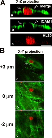
Figure S1. ICAM1-positive membrane sheets surround an adhering and migrating HL60 cell. (A) Differentiated HL60 cells were stained red with membrane dye FM1-43FX (Invitrogen) before the experiment, placed in the upper compartment of a transwell system, and allowed to migrate to 50 ng/ml SDF-1 for 1 h. HUVECs were grown on transwell filters, treated with TNF-α overnight, fixed, and processed for confocal imaging as described in Materials and methods. The top panel illustrates an x-z projection of an HL60 cell in red (a and c). ICAM1 is stained in green (a and b) and surrounds the migrating cell (arrowheads in a and b). (B) X-Y projection of a migrating HL60 cell in red and ICAM1 in green from apical (a; +3 µm) to midsection (b; +0 µm) to baso-lateral focus plane (c; -2 µm). Note the ICAM1-positive ring (arrowhead; a) around the HL60 cell in the apical focal plane. Bars, 10 µm.