[View Larger Version of this Image]
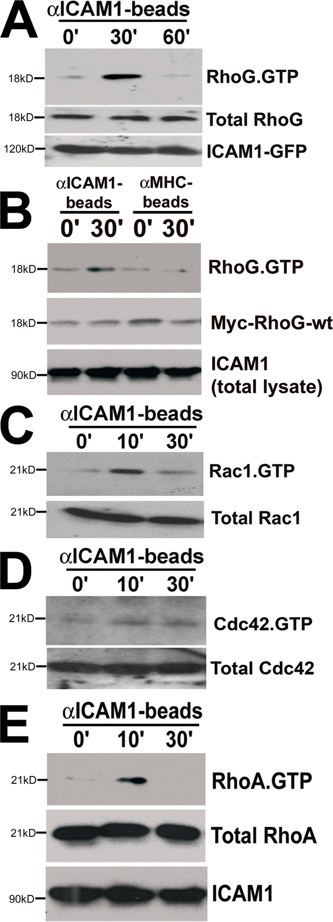
Figure S3. ICAM1 engagement induces the activity of RhoG, Rac1, Cdc42, and RhoA. (A) RhoG was transiently activated downstream from ICAM1 engagement. COS7 cells were transiently transfected with myc-RhoG-wt and ICAM1-GFP. αICAM1 beads were added as described in Materials and methods. Using GST-ELMO, activated RhoG was pulled down from the lysate. Western blot analysis using anti-myc antibodies showed that αICAM1 beads increased RhoG-GTP levels maximally after 30 min and declined after 60 min (top). The middle panel shows protein levels of myc-RhoG-wt in cell lysates. The bottom panel shows ICAM1-GFP expression using anti-GFP antibodies in cell lysates. (B) αMHC class I beads did not activate RhoG in HUVECs. HUVECs were transiently transduced with myc-RhoG-wt and treated with TNF-α. αICAM1 beads or αMHC class I beads were added as described in Materials and methods. Using GST-ELMO, activated RhoG was pulled down from the lysate. Western blot analysis using anti-myc antibodies showed that αICAM1 beads increased RhoG-GTP levels but that αMHC class I beads did not (top). The middle panel shows protein levels of myc-RhoG-wt in cell lysates. The bottom panel shows endogenous ICAM1 expression in cell lysates. (C) Rac1 was transiently activated downstream from ICAM1 engagement. αICAM1 beads were added as described in Materials and methods to TNF-α-treated HUVECs. Using GST-PBD, activated endogenous Rac1 was pulled down from the lysate, and Western blot analysis showed that αICAM1 beads increased Rac1-GTP levels maximally after 10 min and declined after 30 min (top). The bottom panel shows protein levels of Rac1 in cell lysates. (D) Cdc42 was activated downstream from ICAM1 engagement. αICAM1 beads were added as described in Materials and methods to TNF-α-treated HUVECs. Using GST-PBD, activated endogenous Cdc42 was pulled down from the lysate, and Western blot analysis showed that αICAM1 beads increased Cdc42-GTP levels at 10 and 30 min (top). The bottom panel shows protein levels of Cdc42 in cell lysates. (E) RhoA was transiently activated downstream from ICAM1 engagement. αICAM1 beads were added as described in Materials and methods to TNF-α-treated HUVECs. Using GST-Rhotekin, activated endogenous RhoA was pulled down from the lysate. Western blot analysis showed that αICAM1 beads increased RhoA-GTP levels at 10 min, and this declined at 30 min (top). The middle panel shows protein levels of RhoA in cell lysates. The bottom panel shows endogenous ICAM1 expression in cell lysates.