[View Larger Version of this Image]
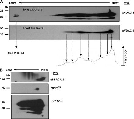
Figure S1. Proteomic analysis of molecular components of the MAM fraction. (A) High-resolution Blue native and SDS-PAGE 2D separation of VDAC1-containing native complexes in the MAM fraction. The native MAM fraction was solubilized with 1 M aminocaproic acid and 2% dodecylmaltoside combined with 5% Serva Blue G and separated on a 4% acrylamide capillary gel in the first dimension. The capillary gel was incubated with a dissociating solution (1% SDS and 1% mercaptoethanol) stacked over a 10% SDS-PA gel and separated; the proteins were then immunoblotted against VDAC1 (1:5,000; Calbiochem; monoclonal anti-porin 31HL human). Long exposure reveals the whole distribution of VDAC1 (top), and densitometry analysis of the short exposure film (bottom) shows the separate VDAC-containing complexes as overlapping peaks (arrows: solid line, major complexes; dotted line, minor complexes). (B) Localization of the SERCA2 protein on a Blue native and SDS-PAGE 2D immunoblot. 2D separation was performed as described in A, and proteins were immunoblotted against VDAC1, grp75 (1:500; rabbit polyclonal; Santa Cruz Biotechnology, Inc.) and SERCA2 (1:500; goat polyclonal; Santa Cruz Biotechnology, Inc.). SERCA2 colocalizes with high molecular weight VDAC1-containing complexes, whereas it has only a partially overlapping spot with grp75, in contrast to the IP3R (Fig. 1).