[View Larger Version of this Image]
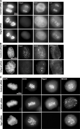
Figure S3. Characterization of cells depleted of hSgo2 by siRNA. (A) Anaphase cells transfected with control (top row) and hSgo2 siRNA (middle and bottom rows) were stained with hSgo2, Mad1, and ACA. The middle row shows a single optical section, and the bottom row shows the maximum projection of the same cell. (B) Prophase cells transfected with control and hSgo2 siRNAs were costained for hSgo2, MCAK, and CENP-E. Chromosomes were stained with DAPI. The arrowhead denotes the centrosomes. Prophase was confirmed by DAPI and the lack of CENP-E at kinetochores. (C) Centromeric localization of MCAK is independent of Sgo1. Cells transfected with control, MCAK, and Sgo1 siRNAs were costained for ACA, Sgo1, and MCAK.