[View Larger Version of this Image]
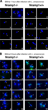
Figure S1. Nramp1-dependent expression of LIT1 by intracellular L. amazonensis. Randomly acquired, independent microscopic fields showing immunofluorescence of C57BL/10ScSn (Nramp1-/-) or B10.L-Lsh (Nramp1+/+) BMmø after 1 (A) or 6 (B) h of infection with L. amazonensis axenic amastigotes. LIT1 is detected after 6 h of infection in Nramp1+/+ BMmø, whereas expression levels remain low in Nramp1-/- BMmø at 1 and 6 h after infection. Antibodies to LIT1 are labeled in green, and the host cell and parasite’s DNA are stained in blue (DAPI). Arrows point to infected macrophages. The images were acquired and enhanced for contrast under identical conditions.