[View Larger Version of this Image]
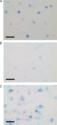
Figure S1. Immunohistochemical analysis of Ig-secreting B-1 cells. Representative photographs for immunohistochemical analysis for cytoplasmic Ig+CD19+/+prdm1Flox/Flox B-1 (A), CD19Cre/+prdm1Flox/Flox B-1 (B), and 4-d LPS-treated B-2 splenocyte (C) cell culture from two independent experiments. Total cells per field were determined by phase contrast. Bar, 50 µm.