[View Larger Version of this Image]
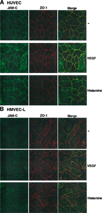
Figure S1. JAM-C localization in macrovascular and microvascular endothelial cells. Analysis of the interendothelial contacts of HUVECs (A) and HMVEC-L (B) was performed. Representative immunofluorescence of quiescent cells (-) or cells that were incubated in the presence of VEGF (1 h, 50 ng/ml) or histamine (1 h, 50 µM). The distribution of JAM-C and ZO-1 is shown. Double stained images were merged to analyze colocalization. Thus, whereas in HUVEC JAM-C is constitutively at interendothelial contacts, in microvascular HMVEC-L JAM-C is localized intracellularly and recruited to the junctions upon stimulation with VEGF or histamine.