[View Larger Version of this Image]
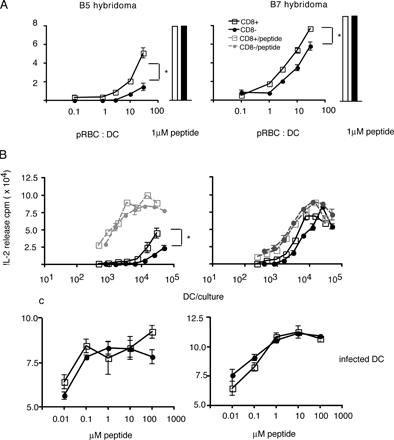
Figure S3. CD8+ DCs from uninfected mice presented MSP1 peptides more efficiently than CD8- DCs in vitro. (A) CD11c+ DCs enriched from spleens of uninfected BALB/c mice were sorted into CD8+ (open squares) or CD8- DCs (closed circles) and cultured with 2 x 10⁴ B5 or B7 hybridoma cells for 24 h in the presence of pRBCs (30:1 schizonts/DC), and IL-2 production was measured as described in Materials and methods. As a control for the presentation capacity, the sorted DCs were cultured in the presence of peptide; bar graphs show the response of the T cell hybridomas cultured with 5 x 10 3 CD8+ (white bars) and CD8- (black bars) DCs and 1 µM of the relevant peptide. (B) Different numbers of sorted DCs were cocultured with the T cell hybridomas in the presence of 30:1 schizonts/DC (solid lines) or 1 µM peptide (dotted lines). The data represent means and SEM (>10%; error bars) of triplicate samples. Statistical analysis was performed as described in Materials and methods. This figure shows one out of three experiments performed. (C) CD11c+ DCs enriched from spleens of infected BALB/c mice were sorted into CD8+ DCs (open squares) or CD8- DCs (closed circles) and cultured at 5 x 10³ DCs/well with 2 x 10⁴ B5 or B7 hybridoma cells for 24 h in the presence of different concentrations of B5 and B7 peptide, and IL-2 production was measured as described in Materials and methods. *, P = 0.05-0.01.