[View Larger Version of this Image]
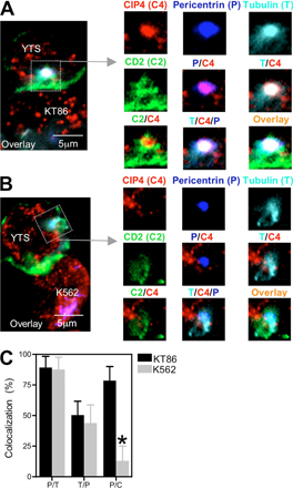
Figure S2. Colocalization in the region of the NK cell MTOC in cytolytic and noncytolytic conjugates. The YTS CD2-GFP cell conjugated with a KT86 target cell (A) or a K562 target cell (B) from Fig. 2 was analyzed using a 5-µm2 region (box) highlighting the MTOC (left). An overlay of all fluorescence is shown and includes CD2-GFP (green), pericentrin (blue), α-tubulin (cyan), and CIP4 (red). The fluorescence contained within the 5-µm2 region was enlarged ∼1.5-fold and is shown (right). The individual images depict the separate fluorescence for CIP4 (C4), pericentrin (P), α-tubulin (T), and CD2 (C2), as well as the merged fluorescence of pericentrin and CIP4 (P/C4); α-tubulin and CIP4 (T/C4); CD2 and CIP4 (C2/C4); α-tubulin, CIP4, and pericentrin (T/C4/P); and an overlay of all four fluorescent signals (overlay). (C) Colocalization within the 5-µm2 region demonstrating the percentage of pericentrin fluorescent pixels that contain α-tubulin fluorescence (P/T), α-tubulin fluorescent pixels that contain pericentrin fluorescence (T/P), and pericentrin fluorescent pixels that contain CIP4 fluorescence (P/T). Almost all of the pericentrin fluorescent pixels also contained α-tubulin fluorescence, whereas approximately half of the α-tubulin fluorescent pixels contained pericentrin. The percentage of colocalization was measured separately in YTS CD2-GFP cells conjugated with KT86 (black bars; n = 10) or K562 (gray bars; n = 10) target cells. Bars represent the mean, and error bars show the SD. The only significant difference of colocalized fluorescence within the specified region in YTS CD2-GFP cells between those conjugated with KT86 and K562 target cells was that there was less colocalization between pericentrin and CIP4 in those conjugated with K562 cells. *, P