[View Larger Version of this Image]
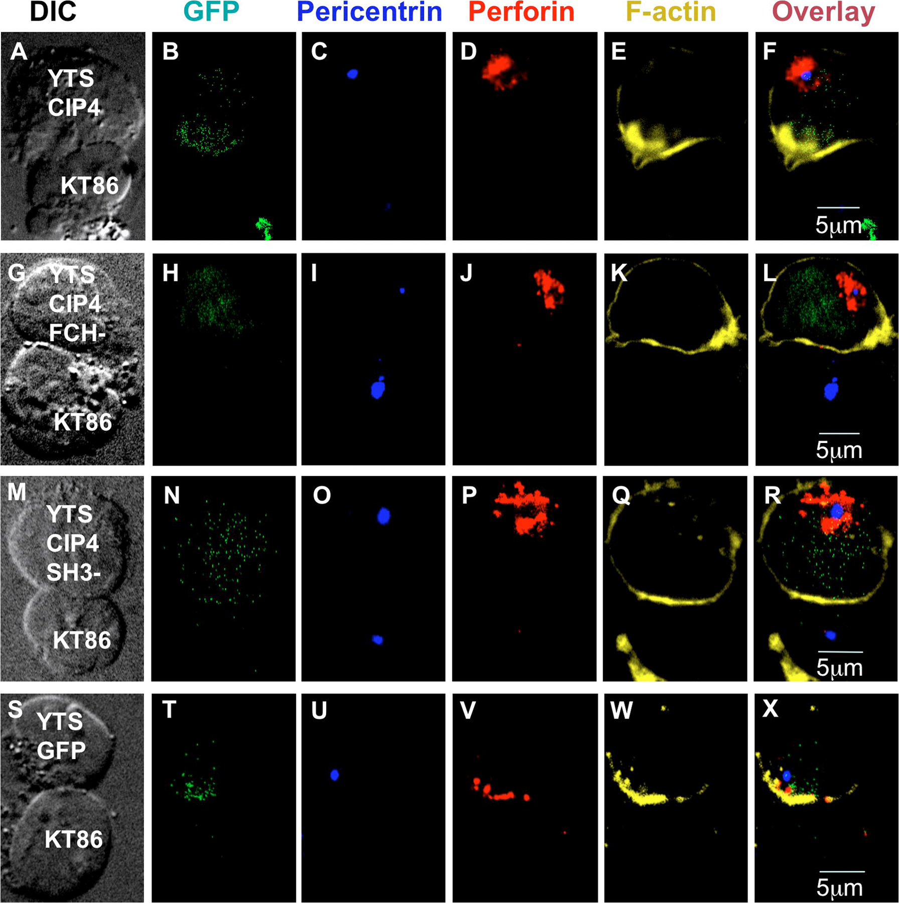
Figure S3.Effect of CIP4 overexpression on perforin localization and the IS in YTS cells conjugated with susceptible target cells. The IS formed between a YTS cell overexpressing WT CIP4 (A-F), CIP4 deleted of the FCH domain (G-L), CIP4 deleted of the SH3 domain (M-R), or GFP only (S-X), and a KT86 target cell is shown. All YTS cells express GFP, which in the cells expressing CIP4 is derived from the bicistronic expression of GFP within the transduced construct. Individual images show DIC microscopy (A, G, M, and S) or confocal microscopy demonstrating fluorescence for GFP (B, H, N, and T), pericentrin (C, I, O, and U), perforin (D, J, P, and V), F-actin (E, K, Q, and W), and an overlay of all fluorescent channels (F, L, R, and X). Each image series displays accumulation of F-actin at the IS but a lack of perforin and MTOC polarization only in those overexpressing CIP4.