[View Larger Version of this Image]
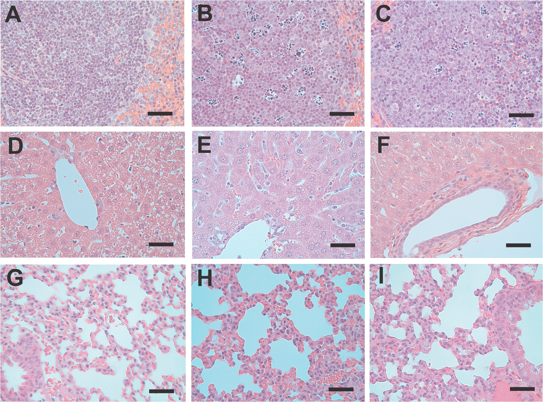
Figure S1. Histopathology of LPS-challenged mice with and without APC treatment. Hematoxylin and eosin-stained sections of spleen (A-C), liver (D-F), and lung (G-I) prepared from control mice (A, D, and G), mice 24 h after LPS challenge/PBS infusion (B, E, and H), and LPS-challenged mice treated with APC (C, F, and I). Bars, 50 µm.