[View Larger Version of this Image]
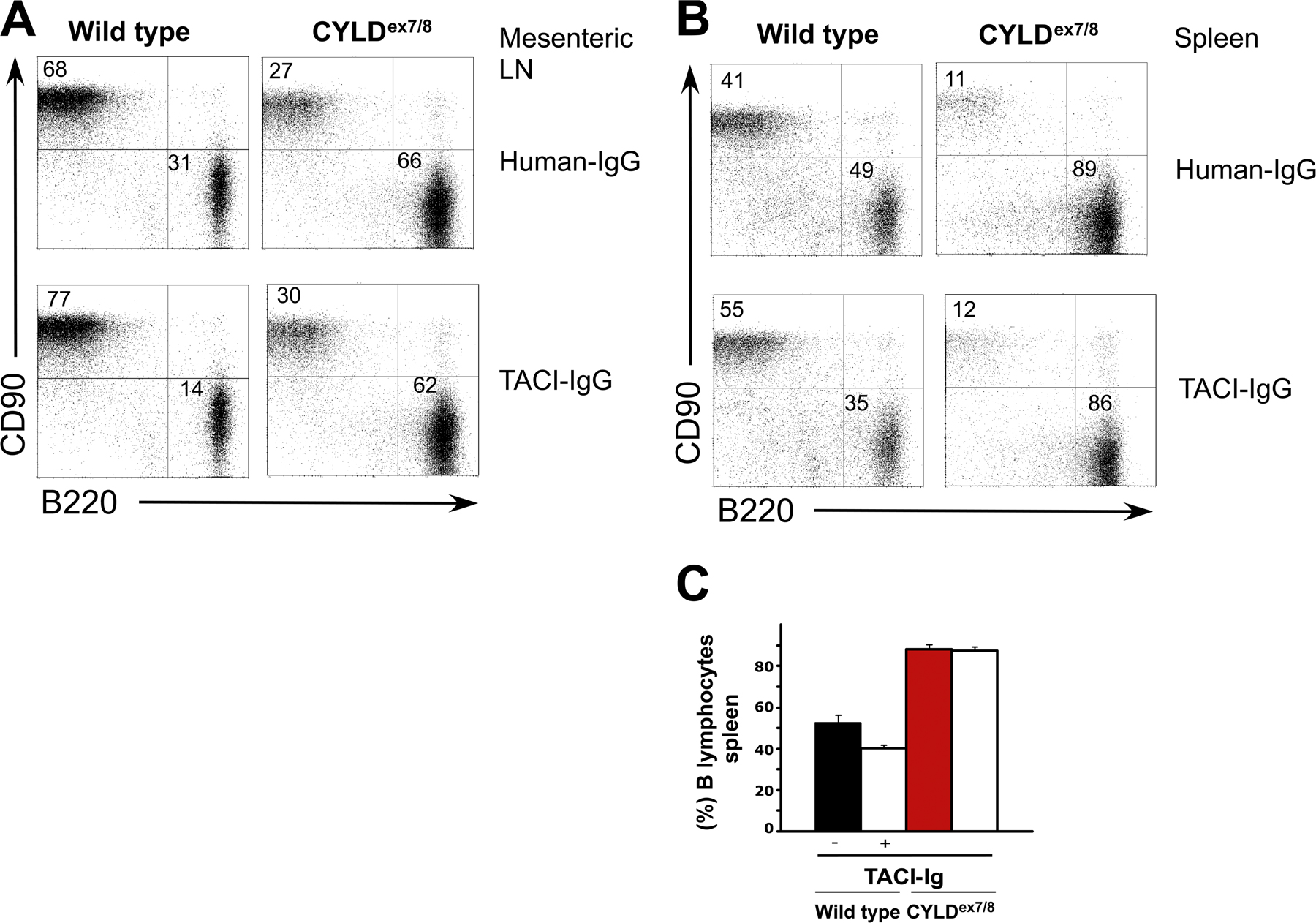
Figure S4. TACI-Ig treatment of CYLDex7/8 and control mice. CYLDex7/8 and WT mice (n = 3) were treated twice a week for 1 wk with 20 µg of TACI-Ig or Hu-Ig control protein. (A) FACS analysis of mesenteric lymphocytes prepared from WT and CYLDex7/8 mice treated with human-Ig or TACI-Ig using B220 and CD90 antibodies to distinguish B and T cell populations. (B) FACS analysis of splenic cells prepared from WT and CYLDex7/8 mice treated with human-Ig or TACI-Ig using B220 and CD90 antibodies to distinguish B and T cell populations. (C) Comparison of percentages of splenic B cells from CYLDex7/8 and WT mice treated with control Hu-Ig (-) and TACI-Ig (+) are shown. Values are the mean ± the SEM.