[View Larger Version of this Image]
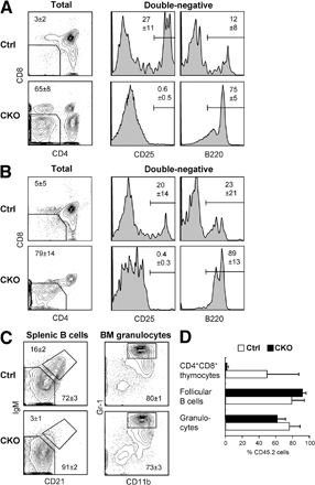
Figure S1. Lymphocyte development after RBP-J deletion in Mx1-Cre CKO mice. Control (RBP-Jfl/fl) or CKO (RBP-Jfl/fl Mx1-Cre+) mice were injected with poly(I):(C) to induce the deletion of RBP-J in the BM. (A) T cell development in the thymus. 3 wk after the deletion, all CKO mice showed very small thymi containing mostly double-negative (DN; CD4-CD8-) cells. Although control DN thymocytes included a large proportion of CD25high pre-T cells and few B220+ B cells, CKO DN cells lacked pre-T cells and consisted primarily of B cells (including a CD25low population corresponding to pre-B cells developing in situ). Mean percentages ± SD of three animals are shown. Splenic MZ B cells showed only a modest reduction, likely because of to their long lifespan (not depicted). (B-D) BM from control or CKO (CD45.2+) mice was transferred into irradiated CD45.1+ recipients, and their lymphoid organs were analyzed 4-5 wk afterward. (B) Donor T cell development in the thymi of chimeric mice. Thymocytes were gated on CD45.2+ donor-derived cells and analyzed as in A. Mean percentages ± SD of six animals are shown. Note a nearly complete block of T cell development and switch to B cell development. (C) B cell development and granulopoiesis in the CD45.2+ donor-derived cells. Shown are follicular (IgMlow CD21-) and MZ (IgMhigh CD21+) subsets of splenic B220+ AA4.1- B cells and BM granulocytes (CD11b+ Gr-1high subset of SSChigh BM cells). (D) The contribution of donor BM to cell lineages in the recipient mice. Shown are mean percentages ± SD of donor-derived (CD45.2+) cells in the indicated cell populations. Note the efficient CKO BM contribution to B cell development and granulopoiesis but not to T cell development.