[View Larger Version of this Image]
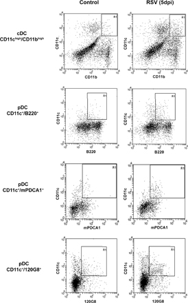
Figure S1. Analysis of CD11chigh/CD11bhigh, CD11c+/B220+, CD11c+/mPDCA1+, and CD11c+/120G8+ DCs. Dot plots showing single cell suspensions of lungs stained for the presence of cDCs (CD11chigh/CD11bhigh) and pDCs (CD11c+/B220+, CD11c+/mPDCA1+ or CD11c+/120G8+) at 0 or 5 dpi. Cells in region R1 were enumerated and depicted in Fig. 1.