[View Larger Version of this Image]
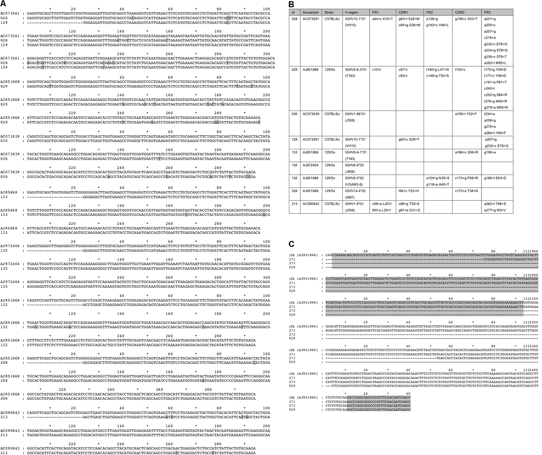
Figure S1. cDNA and genomic sequences of H chain IgG from L−/− mice. (A) Aligned VH cDNA sequences listed in Table I. Matches to the closest VH region were performed using IMGT/V-QUEST (Giudicelli, V., D. Chaume, and M.P. Lefranc. 2004. Nucl. Acids Res. 32:W435–W440). Kabat numbering was performed. Shading indicates differences. (B) Accession numbers, source, and list of mutational alterations. (C) Genomic Cγ H chain sequences, identified by the long-range PCR shown in Fig. 6, identified break points within γ2b. The shaded boxes mark exon 1 (CH1; top) and hinge (5′ region; bottom).