[View Larger Version of this Image]
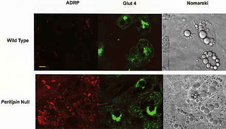
Figure S1. Differentiated fibroblasts from both wt and perilipin-null adipocytes co-stain for ADRP and GLUT4. Fibroblasts from both wt and perilipin-null embryos were differentiated into adipocytes and immunostained for ADRP and GLUT4. Staining for ADRP and GLUT4 is as indicated. Note that the majority of cells reveal prominent perinuclear staining for the glucose transporter. Note also that the adipocytes from the perilipin-null embryos contain prominent lipid droplets that are coated with ADRP. ADRP was visualized with goat anti-murine ADRP followed by donkey anti-goat antibody conjugated to Cy5, and GLUT 4 was visualized with rabbit anti-GLUT 4 followed by donkey anti-rabbit antibody conjugated to FITC. Bar, 10 µm.