[View Larger Version of this Image]
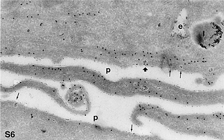
Figure S6. Actin distrubution near the plasma membrane caveolae of SHaPrPC-expressing CHO/30C3 cells. Cells expressing PrPC were incubated with protein A-gold (5 nm, 10-min pulse, and 50-min chase) before fixation. Sections were labeled for actin using the anti-actin antibodies (15-nm gold). Bundles of actin (large gold; as close as 20 nm from caveolae) that are located parallel to the plasma membrane could be seen. Some caveolae (,5%, 20 cells counted, thick arrow) showed actin (large gold) labeling around the protein A-gold (small gold) containing caveolae. e, endocytic structure; l, lysosome; p, plasma membrane. The small arrows are pointing to all protein A-gold-containing caveolae.