Blood, Vol. 110, Issue 9, 3218-3225, November 1, 2007
Effect of presenilins in the apoptosis of thymocytes and homeostasis of CD8+ T cells
Blood Maraver et al. 110: 3218
Supplemental materials for: Maraver et al
Files in this Data Supplement:
- Figure S1. Apoptosis were higher in anti-CD3 mAb-treated control animals than in PS KO animals (JPG, 60.2 KB) -
PBS or anti-CD3 mAb were injected ip in control or PS KO animals and thymii were collected 40h after injection to perform TUNEL analysis in situ. Representative of 2 animals.
- Figure S2. Thymocyte maturation levels in PS KO animals (JPG, 86.7 KB) -
Fresh isolated thymocytes from control or PS KO animals were labeled with fluorescent anti-CD3 and anti-CD24 mAb antibodies. DP cells labeled with anti-CD24 were divided in CD3 negative, low, or high for CD3 expression (A). For CD4 and CD8 SP thymocytes cells were divided in 4 groups according to the CD3 and/or CD24 expression (B and C). Each bar represents the arithmetic mean and standard deviation of 3 animals.
- Figure S3. RORγ expression and NF-κB activity in control or PS KO thymocytes (JPG, 54.5 KB) -
Protein extracts from Control or PS KO thymocytes were used to measure the amount of ROR (A). Nuclear protein extracts were obtained after stimulation (or not) of thymocytes from control or PS KO animals with anti-CD3 mAb, PMA, or TNF-
 , and analyzed by EMSA (B). Representative of 3 animals.
- Figure S4. Activation of p38, ERK, and JNK in thymocytes from control or PS KO animals (JPG, 64.6 KB) -
Protein extracts were obtained after in vitro stimulation (or not) of thymocytes from control or PS KO animals with anti-CD3 mAb for 5 to 20 min. Protein extracts were obtained after each period of time and expression of both active and total p38, JNK, and ERK were visualized by WB. Representative of 3 animals.
- Figure S5. Percentage of CD4 and CD8 cells in Lymph nodes and percentages of TCRγδ T cells in thymii, Lymph nodes, and spleens of control and PS KO animals (JPG, 92.7 KB) -
Lymph node cells (A and B) and/or thymocytes and splenocytes (B) from control or PS KO animals were staining with fluorescent-labeled mAb against CD3, CD4, CD8, TCR
 . Representative of 3 animals.
- Figure S6. Activation markers in peripheral T cells (JPG, 64.9 KB) -
TCR /
/ , CD44, CD62L, CD69, and CD122 expression were evaluated in CD4+ or CD8+ T cells from splenocytes of CD4-Cre (thin lines), PS KO (thicker lines), or Control (full histograms) animals. Representative of 2 tested animals per group.
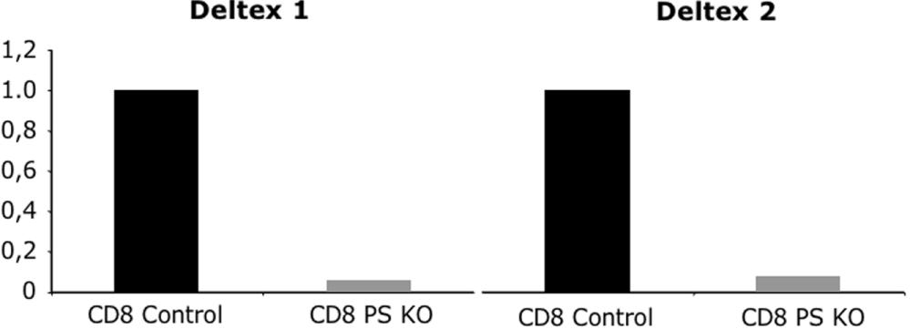
- Figure S7. Expression of Deltex-1/2 genes in CD8 peripheral cells from both control and PS KO animals (JPG, 37.4 KB) -
cDNA from CD8 sorted cells from spleens of Control and PS KO animals were analyzed by real time PCR for Deltex-1/2 genes expression. The expression of PS KO CD8 cells was normalized versus the expression in the Control cells.