[View Larger Version of this Image]
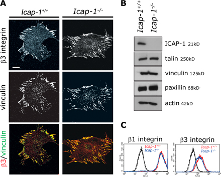
Figure S1. ICAP-1 loss induces no change in the distribution of β3 integrin containing FA and in the expression of adhesion proteins. (A) Confocal images of Icap-1+/+ and Icap-1−/− MEF cells. Cells were cultured overnight on 1 µg/ml FN and processed for immunostaining to visualize β3 integrin and vinculin. (B) ICAP-1 and FA protein expression in Icap-1+/+ and Icap-1−/− MEF cells. An equal amount of protein from cell lysates in radioimmunoprecipitation assay was subjected to Western blotting analysis using either anti–ICAP-1 pAb, antitalin, antivinculin, or antipaxillin mAb. The same membrane has been blotted with anti-actin mAb to control loading. (C) Cell surface analysis of β1- and β3-integrin expression on Icap-1+/+ and Icap-1−/− MEF cells estimated by flow cytometry. (left) Icap-1+/+ (black line and red line) or Icap-1−/− cells (dashed line and blue line) were stained with control antibody (black line) or with anti–β1 MB1.2 mAb. (right) Icap-1+/+ (black and blue lines) or Icap-1−/− (dashed and red lines) cells were stained with control antibody (black line) or with anti–β3 rat mAb. Bar, 20 μm.