Blood, Vol. 111, Issue 4, 1999-2006, February 15, 2008
Prophylactic thrombolysis by thrombin-activated latent prourokinase targeted to PECAM-1 in the pulmonary vasculature
Blood Ding et al. 111: 1999
Supplemental materials for: Ding et al
Files in this Data Supplement:
- Figure S1. Presence of thrombin-cleavage site in the prototype scFv/uPA (JPG, 29.5 KB) -
scFv/uPA was incubated with thrombin and its migration determined on non-reduced SDS-PAGE exactly as described in “Methods” for scFv/uPA-T. Thrombin cleaves scFv/uPA and generates two bands with distinct molecular sizes. In contrast, it was shown previously that plasmin treatment of scFv/uPA does not affect its migration on non-reduced SDS-PAGE gels, but generates two bands that migrate similarly on reduced SDS-PAGE gels.
- Figure S2. Thromboplastin injection increased deposition of fibrin/fibrinogen but not IgG in mouse lungs (JPG, 29.5 KB) -
To test whether the deposition of fibrinogen in the lung is attributable to thrombosis in thromboplastin model, we measured the deposition of radiolabeled IgG (120,000 cpm/mouse) injected intravenously along with reconstituted thromboplastin or with the same volume of PBS in the identical manner to labeled fibrinogen as described in “Methods.” Residual radioactivity in the lung was measured and calculated as percent of injected dose per gram tissue (%ID/g). Thromboplastin injection did not significantly increase the IgG deposition in mouse lungs, whereas pulmonary deposition of radiolabeled fibrinogen was increased ~4-fold (from 5%ID/g to 22%ID/g). This indicates that extravasation of plasma proteins contributes very little to the accretion of radiolabeled fibrinogen seen after injection of thromboplastin. Rather, the deposition of fibrinogen occurs as a result of thrombosis and accrual of fibrin (**, P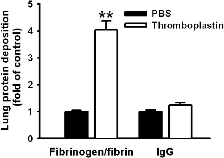
- Figure S3. Endothelium-anchored scFv/uPA-T attenuates pulmonary fibrin deposition in a mouse model of thrombin-induced thrombosis (JPG, 44.2 KB) -
In control mice without thromboplastin injection, 5 ± 0.4% of injected radiolabeled fibrinogen was found in the lungs, reflecting basal level of non-specific retention. In PBS-treated mice injected with thromboplastin, 22 ± 1.8% of the injected radioactivity was deposited per gram of lung tissue, reflecting pulmonary thrombosis. Percent of fibrin deposition in the lungs of animals given scFv/uPA, scFv/uPA-T or scuPA was calculated using the formula: Fibrin deposition (%) = (Residual radioactivitydrug – Residual radioactivityFg) / (Residual radioactivityPBS – Residual radioactivityFg) ×100 (*, P vs. lmw-scuPA; #, P vs. scFv/uPA).
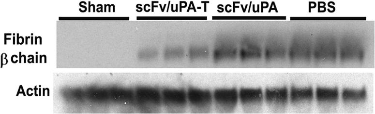
- Figure S4. Lung fibrin deposition in mice treated with uPA formulations and subjected to I/R (JPG, 26.5 KB) -
After mice were injected with the indicated uPA formulations or vehicles and subjected to I/R as described in “Methods,” Western blots were performed using anti-fibrin -chain antibody. The amount of fibrin was quantified and normalized to actin, as described in “Methods.” Representative western blot results are shown. Each lane represents one mouse.