[View Larger Version of this Image]
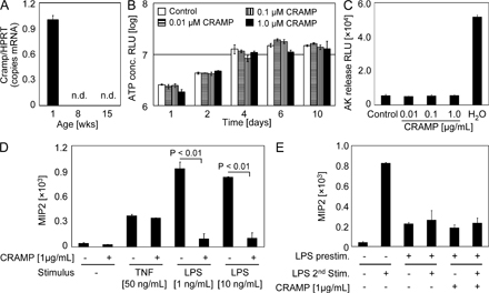
Figure S1. Effect of CRAMP on cell proliferation and LPS recognition by neonate IECs. (A) Quantitative analysis of Cramp mRNA expression in primary IECs isolated from the small intestinal tissue of 1-, 8-, and 15-wk-old mice. Values represent the mean ± SD of gene expression determined in total IECs and indicate the target/housekeeping (Hprt1) gene expression ratio. n.d., not detectable. (B) m-ICcl2 cells seeded at 103 cells per well were incubated in the absence or presence of CRAMP at the indicated concentrations for 10 d, and cell proliferation was determined by ATP quantification after complete cell lysis using the ViaLight Plus kit (Lonza). (C) Confluent m-ICcl2 cells were exposed to the indicated CRAMP concentrations overnight, and adenylate kinase (AK) release in the cell-culture supernatant was determined using the ToxiLight cytotoxicity assay (Lonza) according to the manufacturer’s instructions. (D) Intestinal epithelial m-ICcl2 cells were stimulated with TNF or LPS at the indicated concentrations in the absence or presence of CRAMP. Cellular activation was determined by quantification of macrophage inflammatory protein 2 (MIP2) secretion in the cell-culture supernatant. (E) Intestinal epithelial m-ICcl2 cells were left untreated or prestimulated with 1 µg/ml LPS for 6 h. MIP2 was determined in the cell-culture supernatant after a second stimulation 36 h later with 10 ng/ml LPS in the absence or presence of CRAMP over 6 h. Values represent the mean ± SD.