[View Larger Version of this Image]
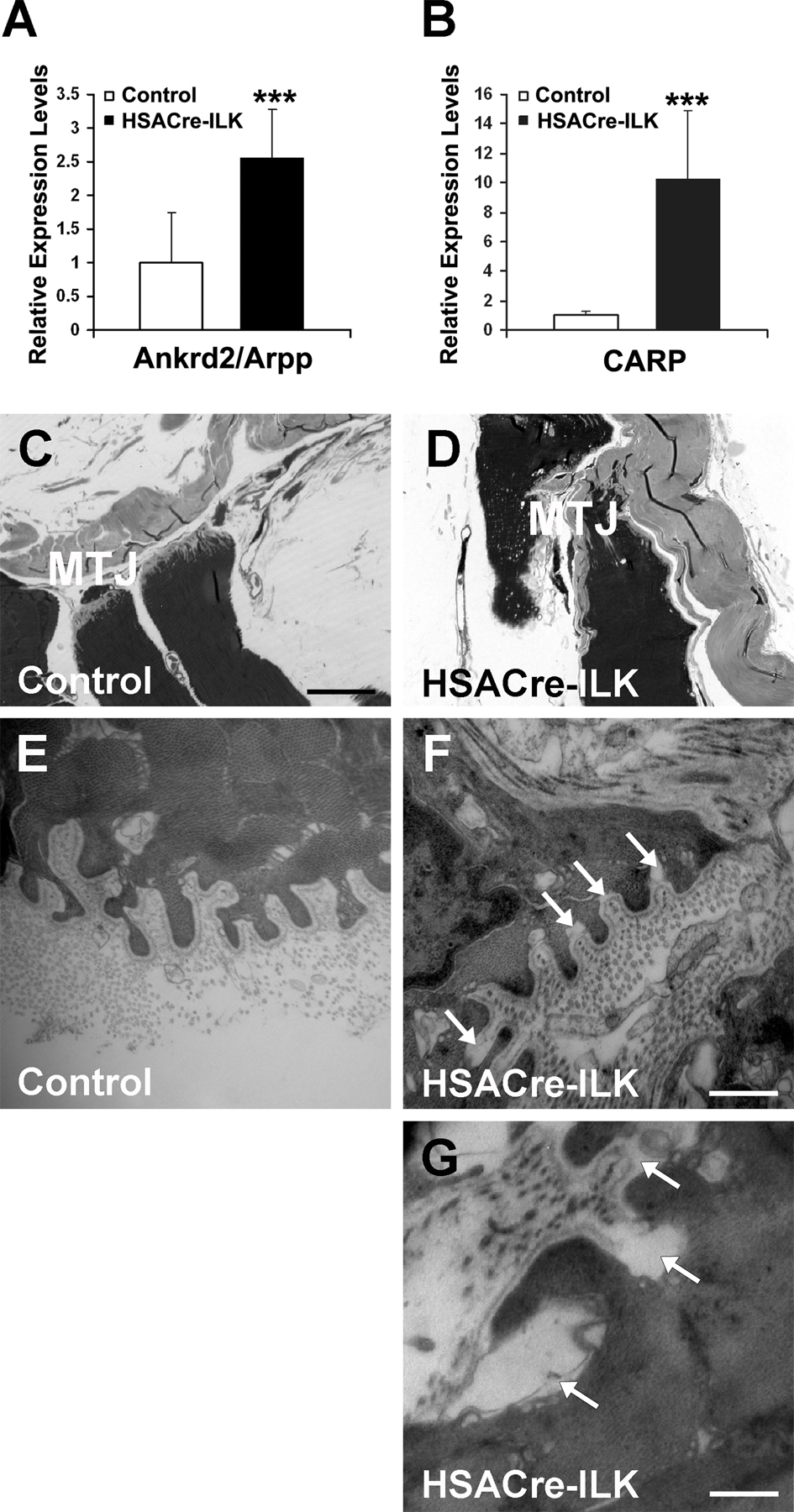
Figure S5. Muscle damage and detachment of BM in trained HSACre-ILK muscle. (A and B) Relative mRNA levels of Ankrd2/Arpp and CARP are shown, measured by quantitative PCR in GC muscle from trained control and HSACre-ILK mice. The levels of both Ankrd2/Arpp and CARP are significantly elevated in HSACre-ILK muscle. Data are expressed as mean ± SD (n = 3; ***, P < 0.001). (C–G) Light (C and D) and electron (E–G) micrographs of skeletal muscle fibers of control (C and E) and HSACre-ILK (D, F, and G) mice. (C) Normal MTJ with regular short interdigitations. (D) HSACre-ILK MTJ lost the typical shape and shows cytoplasmic processes and invaginations with variable size and shape. (E–G) The fingerlike folds of MTJ are covered by a closely attached BM in trained control (E) muscle. The BM (arrows) is frequently detached at less affected fingerlike folds (F) and almost completely between irregularly shaped cytoplasmic processes of severely affected MTJ in HSACre-ILK (G) mice. Bars: (C and D) 35 µm; (E and F) 900nm; (G ) 1.4 µm.