[View Larger Version of this Image]
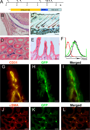
Figure S3. Generation and characterization of ColVI-Cre mice. (A) Representation of transgene construct. The SphI fragment contained 7.5 kb of the Y-flanking sequence of the gene, the first two exons, and the first and part of the second intron, and included a PstI fragment extending from base +41 to base −1344 from the transcription start site that was ligated to the Cre-hGH cassette. The SalI-NotI fragment was injected into the pronuclei of one-cell-stage embryos (C57BL/6 × CBA), and surviving embryos were implanted into pseudopregnant foster mothers. B, BamHI; P, Psti; S, SphI. (B) Functional analysis of the ColVI-Cre ROSA26flx/+ mouse. X-gal staining in whole joints. Bar, 25 μm. (C) X-gal staining on skin-section keratinocytes and skin mesenchyma. Bar, 10 μm. (D and E) X-gal staining on ileum sections. Bars: (D) 30 μm; (E) 80 μm. (F) Flow cytometric analysis for quantification of recombination efficiency. Single-cell suspensions were examined for β-galactosidase expression using fluorescein di-β-D-galactopyranoside (FDG; Invitrogen), according to manufacturer’s instructions. The efficiency of recombination in ColVI-Cre ROSA26flx/+(green line) is compared with ROSA26dfx/+ (black line) and ROSA26flx/+ (red line) control samples. (G–I) CD31 staining of ileum slides from ColVI-Cre Z/EG mice. No colocalization of enhanced GFP with the endothelium marker CD31 (BD Biosciences). (J–L) αSMA (neomarkers) staining of ileum slides from ColVI-Cre Z/EG mice. Bars, 40 μm.