[View Larger Version of this Image]
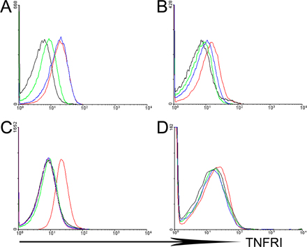
Figure S4. TNFRI expression on mesenchymal and nonmesenchymal tissues. Flow cytometric analysis for the detection of TNFRI expression in ColVI-Cre TnfRIflxneo/flxneo mice. SFs (A) and IMFs (B) were derived from pooled cultures of two mice per group, whereas splenocytes (C) and BM-derived macrophages (D) were derived from the individual mice used for SF and IMF cultures, respectively. In brief, 2–5 × 10⁶ cells per experimental animal per group were stained with hamster anti–mouse TNFRI antibody (BD Biosciences), followed by secondary antibody (goat biotinylated anti–hamster IgG; Vector laboratories) and streptavidin-PE (BD Biosciences). The identification of cell populations was performed using specific cell markers for myeloid cells (CD11b-FITC; BD Biosciences) or fibroblasts (CD90.2-FITC; eBioscience). Histograms shown were gated on CD90+ cells or CD11hi cells for SF/IMF and myeloid cells from spleen/BM-derived macrophages, respectively. WT (red line), TnfRI−/− (black line), TnfRIflxneo/flxneo (green line), and ColVI-Cre TnfRIflxneo/flxneo (blue line) are shown.